Discover More with Cryo-Fluorescence Tomography
Transformative 3D Imaging to Monitor Drug PK/PD
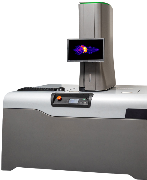
Contact UsCryo-Fluorescence Tomography (CFT) is a transformative 3D approach to imaging drug distribution and protein expression in whole animals. EMIT Imaging offers both instrumentation and services via our imaging platform, XerraTM, a high resolution and high sensitivity preclinical imaging tool designed to advance discoveries in biological and drug research. CFT can be easily integrated into your existing in vivo imaging workflows. See what you’re missing with standard 2D in vivo fluorescence imaging techniques and utilize CFT to better visualize:
- Whole-body drug distribution and delivery
- Screening candidate drugs and delivery systems
- Whole-body therapeutic protein expression
- Multiplexed co-localization of drug with targets
- On and off target tissue identification
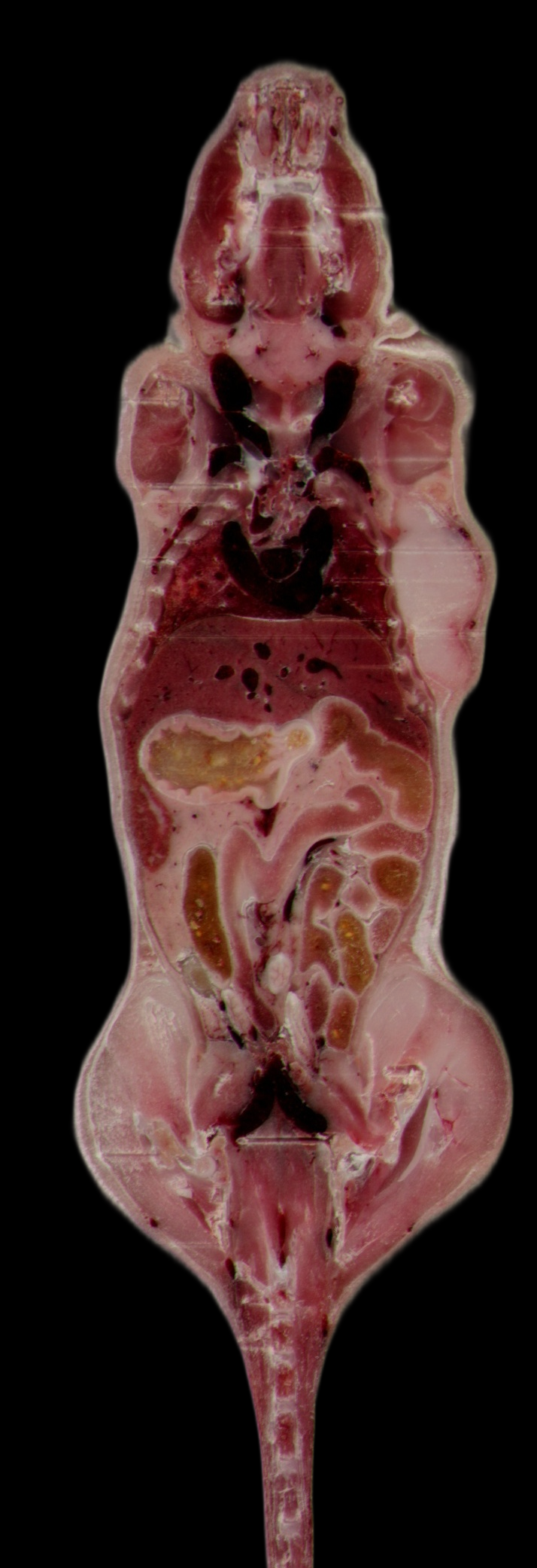
Fibroblast-activation protein: Whole Body Biodistribution Copyright 2023 Ratio Therapeutics Inc., used with permission

Benefits of Cryo-Fluorescence Tomography
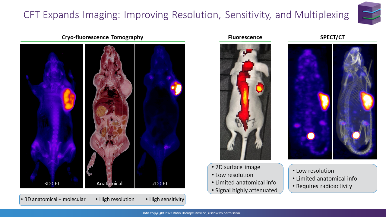
CFT expands preclinical imaging capabilities by bridging the gap between cellular microscopy (imaging small tissue volumes at high resolution) and current whole-body techniques such as MRI, PET, and SPECT (imaging larger tissue volumes with lower resolution and sensitivity). CFT also helps guide technologies, including pathology and proteomics to evaluate all relevant tissues and better develop a complete picture of disease and treatment.
How CFT Works
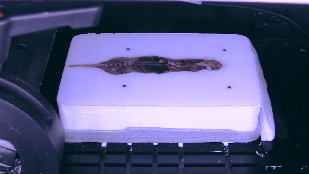
Step 1: Preparation
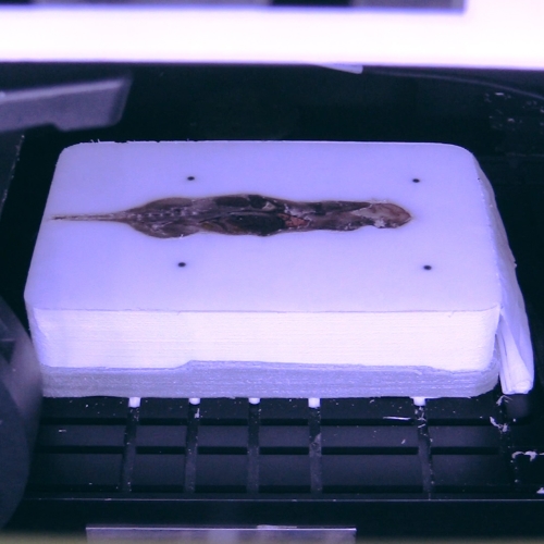
Step 2: Image + Section
Step 3: Output
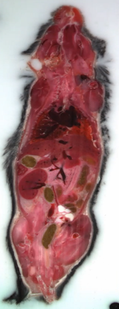
3D image stacks are generated. White light and fluorescence data can be overlaid.
CFT Instrumentation & Services
Instrumentation
Services
EMIT Imaging offers CFT fee-for-service research performed in-house, at our state-of-the-art labs in Boston, MA and Baltimore, MD. Simply freeze samples, ship them directly to us, and let EMIT handle the rest.
Application Areas
Drug Discovery & Delivery
CFT is an important tool to monitor drug distribution, pharmacokinetic, and pharmacodynamic studies.
Oncology & Immunotherapy
CFT can play a key role for studying tumor models including microenvironments, tumor heterogeneity, metastatic spread, and expression of specific biomarkers.
Gene & Cell Therapy
(CFT) and Xerra™, a 3D imaging technology allows whole-body detection and visualization of vector biodistribution and vector-mediated gene expression, in high resolution and with high sensitivity.
Neuroscience
The 3D CFT maps of fluorescence reporters can aid in visualization of cell tracking, drug delivery, and pharmacodynamics in the brain.