Enhancing Preclinical PET/SPECT Imaging
with Cryo-Fluorescence Tomography
Positron Emission Tomography (PET) and Single Photon Emission Computed Tomography (SPECT) are essential in vivo molecular imaging techniques offering spatial and functional insights into biological processes. A huge advantage of nuclear imaging is that it is highly translational and provides quantitative and longitudinal information. Despite its expansive use in the preclinical and clinical spaces, each has inherent spatial resolution limitations, that can hinder precise signal localization, particularly for heterogeneous tissues or small lesions. These considerations are key drivers supporting the need for complementary imaging technologies.
This technical note describes Cryo-Fluorescence Tomography (CFT) as an advanced ex vivo 3D imaging solution. CFT provides high-resolution (20-55 µm pixels) anatomical and molecular fluorescence data from cryo-preserved whole animals and organs. The automated serial sectioning and block-face imaging workflow co-registers volumetric datasets. Key capabilities include single-digit nanomolar sensitivity, resolution down to 20 µm pixel resolution, fluorescence multiplexing, volumetric analysis, and the ability to conduct high-throughput studies. EMIT Imaging’s CFT Instrumentation & Services bridge the gap between macroscopic in vivo scans and microscopic tissue imaging, increasing confidence in research outcomes. CFT can be adopted as an upstream screening tool or downstream as a method to validate in vivo PET/SPECT findings.
The Pros & Cons of Nuclear Imaging Techniques
- Molecular Sensitivity: Capable of detecting picomolar concentrations of radiotracers, enabling visualization of receptor-ligand interactions, enzyme activity, and metabolic flux in vivo
- Quantitative Imaging: that provides measurable biodistribution data, a valuable component when submitting to regulatory bodies
- Whole-body imaging: provides systemic biodistribution information with multi-organ tracking of agents
- Clinical Translatability: the use of FDA-approved or investigational radiotracers assists with translation from pre-clinical research to clinical trials
- Multi-isotope & dual-tracer compatibility: the hybridized use of PET/SPECT or dual-isotope SPECT imaging allows for simultaneous or sequential imaging of multiple biological processes in a single study
- Suitability for longitudinal studies: supporting imaging in the same subjects across a span of time
Despite this method’s many benefits to the research community, it is not without its complexities, which include:
- Radiation safety and compliance demands: Requires controlled-access facilities, dosimetry protocols, and compliance with NRC, IACUC, and FDA regulations or other equivalent regulatory agencies
- High capital and operational expenditure: Requires investment in cyclotron, radiopharmacy or commercial isotope access, equipment and infrastructure, and trained nuclear medicine personnel
- Spatial Resolution Limitations: Typically limited to 0.5-1.0 mm for preclinical systems due to the decay process
- Short physical half-lives of PET isotopes: Isotopes like ¹⁸F and ¹¹C require time-sensitive synthesis and imaging workflows
- Limited throughput and physiological constraints: Long scan times, anesthesia requirements, and animal handling reduce throughput and may alter tracer kinetics
While PET and SPECT are highly effective for tracking radiolabeled compounds in vivo, their limited spatial resolution can hinder precise anatomical localization-especially when studying small lesions, heterogeneous tissue structures, or evaluating off-target effects. Without clear anatomical detail, it becomes difficult to determine whether observed signals originate from intended targets or nearby tissue, introducing ambiguity into biodistribution data and complicating downstream analysis and interpretation.
Overcoming the Limitations of PET & SPECT with Cryo-Fluorescence Tomography
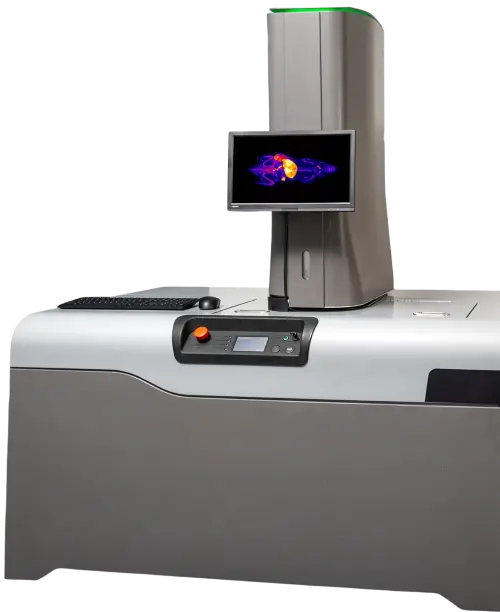
EMIT Imaging is the manufacturer of the Xerra Cryo-Fluorescence Tomography Imaging Platform. CFT involves the cryo-preservation of an entire animal or isolated tissues with the system accommodating multiple organs and up to 10 vertical mice at a time. The Xerra system works by automatically sectioning the frozen sample at thicknesses ranging 20-55 µm. After each section is removed, the block face is imaged, capturing anatomical and fluorescence images. The molecular signals are derived from fluorescent probes, labeled drugs, and reporter gene expression. These sequential 2D images are then reconstructed to generate a high-resolution volumetric dataset.
Key Capabilities and of Cryo-Fluorescence Tomography:
- High-Resolution: Achieve pixel resolutions down to 20 µm, providing sub-organ level detail that surpasses standard in vivo imaging modalities, allowing for enhanced visualization of biodistribution.
- High-Sensitivity: Detect fluorescent signals in the nanomolar range, offering sensitivity that is beneficial for preclinical applications and low-signal targets.
- Volumetric Data: Generate comprehensive 3D datasets, enabling visualization of probe or drug distribution throughout entire organs or even whole small animals in their complete anatomical context.
- Image Co-registration: Automatically capture white light anatomical images that are co-registered with the molecular fluorescence data for each section, allowing for precise co-localization of signal
- Multiplexing Capabilities: The Xerra system is equipped with multiple lasers and filter sets, allowing for the simultaneous detection and differentiation of multiple fluorophores. This enables the study of complex biological interactions, co-localization of different molecular species, or the use of anatomical markers alongside probes of interest.
- Operational Efficiency and Safety: When used for early-stage screening as an alternative to nuclear imaging, CFT allows researchers to avoid the complexities and costs associated with radioactive materials. PET and SPECT rely on radiotracers, which often involve logistical challenges such as the need for on-site production (for short-lived isotopes), complex radiochemistry, and strict timing due to radioactive decay. By using fluorescently labeled molecules, CFT enables whole-body biodistribution assessment without these constraints – delaying the need for radiotracer-based imaging until drug candidates are further optimized.
These capabilities make CFT a powerful tool for increasing confidence in research conclusions. High-resolution ex vivo validation strengthens data interpretation and provides complimentary support for in vivo PET/SPECT findings. With CFT, researchers gain a deeper biological understanding by uncovering nuanced details of probe or drug biodistribution, target engagement specificity, off-target accumulation, and signal heterogeneity that may not be visible through in vivo imaging alone. When paired with striking CFT visuals, these insights become even more compelling for publications and funding proposals. The combination of relative fluorescence quantification and high-impact 3D imagery enhances the clarity and significance of experimental results.
Integrating Cryo-Fluorescence Tomography into the PET/SPECT Workflow
The integration of CFT into a PET/SPECT research workflow is designed to be seamless. CFT can be adopted in two ways: as an upstream screening tool or as an end-point imaging modality.
| Utilization of CFT for Upstream Screening | Utilization of CFT for End-Point Imaging |
| Integrating CFT upstream in the molecular imaging workflow helps streamline candidate selection, avoids the need for use of radioactive materials, and increases confidence in downstream PET/SPECT studies. | Using CFT for endpoint imaging provides detailed anatomical and molecular context, helping validate in vivo results and reveal tissue-level heterogeneity. |
| Step 1: Preparation of pre-screening cohort. Fluorescently labeled therapeutic candidates or reporter genes are administered to a screening subgroup. | Step 1: In vivo PET/SPECT Study, followed by injection of fluorescent probe(s) for CFT. |
| Step 2: Euthanasia of the pre-screening cohort. | Step 2: Euthanasia of the cohort following in vivo study components. |
| Step 3: Whole-animal or individual tissues are cryo-preserved and embedded in EMIT’s proprietary imaging media. | Step 3: Whole-animal or individual tissues are cryo-preserved and embedded in EMIT’s proprietary imaging media. |
| Step 4: The Xerra system performs serial sectioning and anatomical + multi-channel fluorescence imaging. | Step 4: The Xerra system performs serial sectioning and anatomical + multi-channel fluorescence imaging. |
| Step 5: Data analysis and candidate assessment provides whole-body biodistribution screening, target vs. off-target evaluation, and acts as a screening gate. | Step 5: Data analysis & data comparison with PET/SPECT allows for precise signal localization, validation of in vivo hot spots, and multi-modal correlation. |
| Step 6: Candidates with favorable biodistribution are pursued for downstream NM PET/SPECT imaging. |
When used as a screening tool, CFT helps mitigate programmatic, financial, and timeline risks that arise from advancing a failed drug or therapy candidate. It provides a cost-effective solution for R&D teams to efficiently refine potential candidates using whole-body biodistribution insights.
When used as a downstream confirmation method, CFT enables researchers to precisely localize biodistribution patterns – whether expected or not – that may not have been visible in the original scan, impacting decision making for the future direction of the program This clarity empowers confident conclusions and has the advantage of providing compelling visual evidence to strengthen publications and grant proposals.
So we at EMIT Imaging ask: if your science could be more targeted, your workflow more efficient, and your conclusions more definitive – why wouldn’t you take the next step?
Accessing Cryo-Fluorescence Tomography Instrumentation & Services Through EMIT Imaging
EMIT Imaging ensures the scientific research community can easily access Cryo-Fluorescence Tomography in the form of both services and instrumentation. As pioneers of CFT, EMIT Imaging’s team brings a critical technological advancement to market that enhances the validation and comprehensive understanding of PET/SPECT imaging data. By bridging the resolution gap between macroscopic functional imaging and microscopic tissue analysis, CFT delivers co-registered multiplexed datasets with exceptional detail and sensitivity. This enables researchers to precisely localize signals, validate in vivo observations, and gain deeper insights into complex biological systems, ultimately increasing the confidence and impact of their discoveries.
Contact EMIT Imaging
For more detailed specifications on the Xerra Cryo-Fluorescence Imaging Platform, application examples, or to discuss how CFT can be integrated into your specific research workflow, please contact EMIT Imaging at info@emitimaging.com