
Xerra™ imaging platform
A fully automated CFT workflow to advance discoveries in biological and drug research.
Contact UsRun whole-body samples in-house
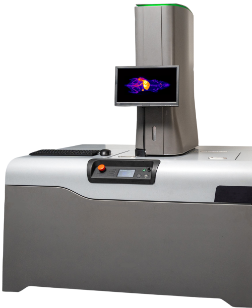
Xerra™ is EMIT Imaging's flagship preclinical imaging platform, available for purchase. Xerra is designed to advance discoveries in biological and drug research and fully automates the CFT workflow, providing 3D images with high-resolution and high-sensitivity. This enables researchers to easily run samples in-house and obtain unmatched visualization of drug distribution and protein expression in whole-animals.

How CFT works
Sample preparation
Cryo-preserve and embed the sample in OCT.
Image + section
High-resolution, high-sensitivity white-light and fluorescence images are acquired. The block is sectioned and 20-55 microns are removed from the surface. The process repeats until the whole sample is sectioned and imaged.
Output
3D image stacks are generated. White-light and fluorescence data can be overlaid.
Research advantages
High resolution
High, near-cellular resolution down to 20 µm.
High sensitivity
Nanomolar sensitivity, comparable to nuclear medicine.
High throughput
Several samples in the same block means a large volume of tissue imaged.
Complementary
Fits into any experiment, complementing tissue and radiology imaging.
Simplified
No fixation, perfusion, clearing, or radiolabeling required.
Multiplexing
6 lasers and 7 filters for multiplexed applications.