Home / Applications / Drug Discovery & Delivery
Application area

Drug discovery & delivery

Monitor on- and off-target expression with resolution and sensitivity.

Overview

CFT for drug discovery and delivery

Cryo-Fluorescence Tomography (CFT) is an ex vivo, volumetric tissue imaging technique used to monitor the pharmacokinetics and pharmacodynamics (PK/PD) of drugs and delivery vehicles with high resolution and sensitivity. Images of the anatomy (white light) are acquired alongside fluorescence images that are displayed in a 3D visualization of the drug and/or delivery vehicle biodistribution.

EMIT Imaging's visualization tools make CFT data easier to explore and collaborate on, with a full range of options to fit different user types and use cases.

Contact us to identify the best visualization tool for your team and receive a demo →
FAP CFT case study thumbnail
Case study
Preferential Tumor Uptake of a FAP-Targeted Therapy
Read the case study →
Talk to a scientist

Optimizing a delivery vehicle?

Send us your study design and someone from our team will follow up.

Research examples
Example · IV administration

AAV-mediated protein expression

AAV9 tropism

Findings: CFT uniquely achieves the required combination of resolution and sensitivity in a whole animal.
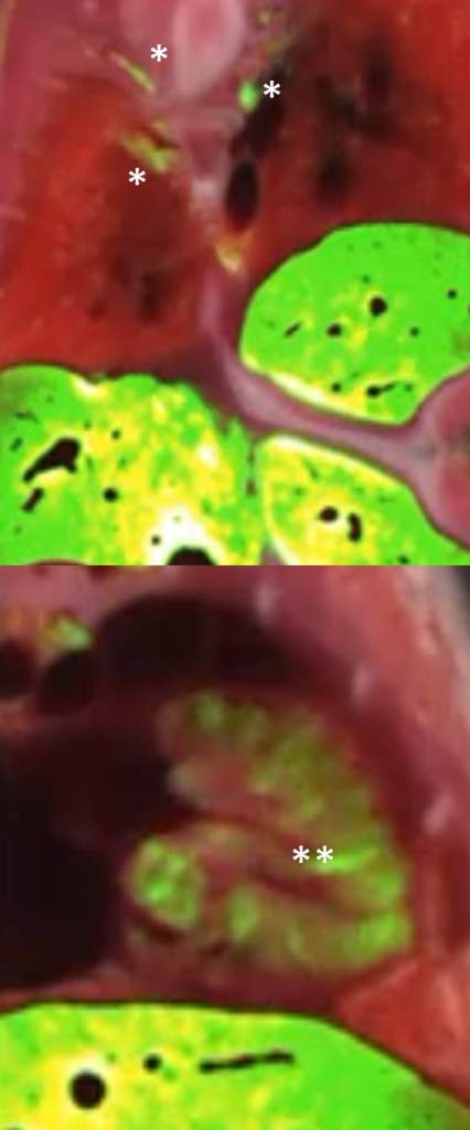
GFP expression at day 15 following IV injection
No expression in the control
Liver is the major organ where GFP expression occurs, but protein expression is observed throughout the body
Note signal in dorsal root ganglion (DRG) and heart, consistent with AAV9 tropism
AAV9 GFP expression, heart and liver — 3D CFT flythrough
AAV9 GFP expression, heart and liver, 3D CFT
AAV9 GFP expression, heart and liver — 3D CFT
Example · intrasplenic cell injection

Visualization of cell intake

Cell tracking

Findings: CFT was used to visualize labeled cell intake into tissue.

Cell tracking by fluorescence labeling is a critical aspect of cell-based therapies, such as CAR-T and regenerative medicine
In preclinical research, cells are administered systemically or directly in target tissues, and localization is evaluated using in vivo or tissue imaging
In this example, proprietary cells were labeled with commercial QTracker 800 (ThermoFisher) and injected into mouse spleen
Primary pellet observed in the spleen and a secondary pellet in the liver
Primary pellet in the spleen
Secondary pellet in the liver
Large formats

CFT application in large animal tissue

NHP · bovine · porcine

Findings: Because Xerra works in large formats, CFT has strong applications for anatomical RGB white-light and fluorescence imaging in large animal tissue, including NHP, bovine, porcine, and more.

Porcine heart: fluorescence Porcine heart: anatomical RGB white-light
White-light
Fluorescence
Porcine heart — drag to compare
Bovine brain: fluorescence Bovine brain: anatomical RGB white-light
White-light
Fluorescence
Bovine brain — drag to compare
White-light of bovine eye
White-light and fluorescence overlay of bovine eye
Fluorescence eye
Fluorescence MIP of bovine eye imaged with CFT
View More Examples in the CFT Image Gallery →
Related applications

Discover More with Cryo-Fluorescence Tomography

Contact Us