Gene & Cell Therapy
Visualization of vector biodistribution and vector-mediated gene expression
Contact UsCFT in Gene and Cell Therapy
New vectors are in development to improve their efficacy, including biodistribution and transduction, while minimizing off-target or safety liabilities. Unbiased visualization of vector biodistribution and subsequent gene transduction in preclinical models are crucial in early-stage research; however, these critical studies are challenging using standard imaging modalities.
See what you’re missing with Cryo-Fluorescence Tomography (CFT) and Xerra™, a 3D imaging technology that allows whole-body detection and visualization of vector biodistribution and vector-mediated gene expression, in high resolution and with high sensitivity.
AAV-mediated Protein Expression Following IV Administration

Example: Whole Body Distribution of Labeled ASO
Intrathecal Injection
Findings: CFT is superior to surface fluorescence due to its depth limitations (signal loss) and poor resolution
Details:
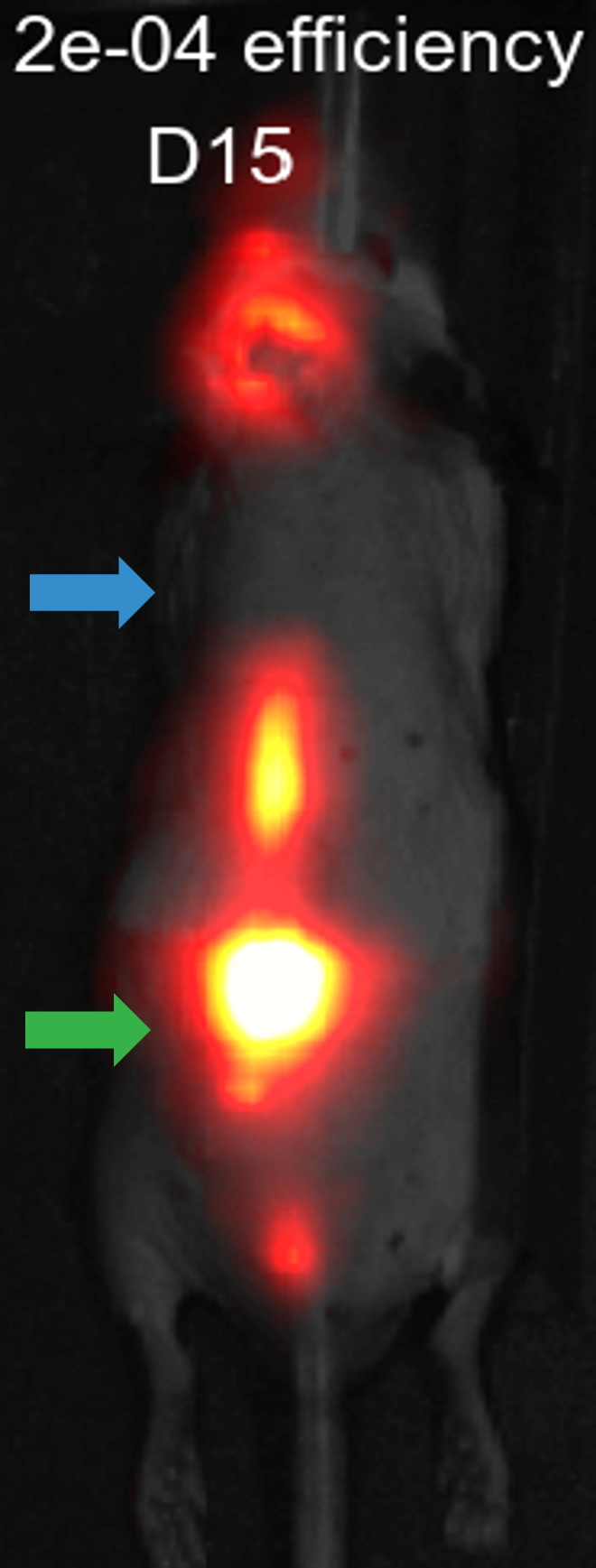
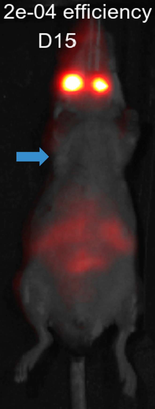
- Cy5-labeled ASO at day 15 following IT injection
- Full visualization of spinal column into the cisterna magna
- CFT detects bioD and clearance: GI, lympathic, nasal turbinate, etc.
- CFT is 3D and high resolution: <50mm versus >1mm in optical and nuclear medicine imaging
–> Signal loss due to depth, note the spine in CFT
-> Site of Injection
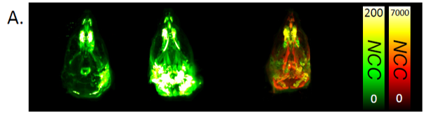
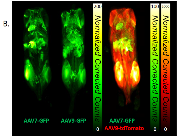
Example: Whole-Body Transduction
AAV7-GFP + AAV9-tdTomato
Findings: CFT visualization of whole-body transduction and expression by AAV9 and AAV7
Details:
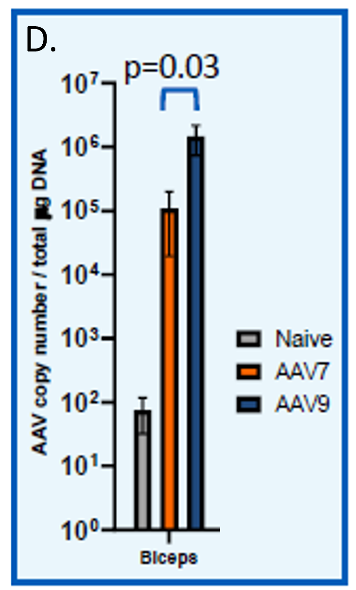
- Representative images of animals administered single vectors or both AAV7.CAG.EGFP and AAV9.CAG.tdTomato. (A,B)
- Representative CFT images of EGFP fluorescence in hind limb muscle (C) and protein copy number by qPCR (D)

Yost et. al, ASGCT 2023
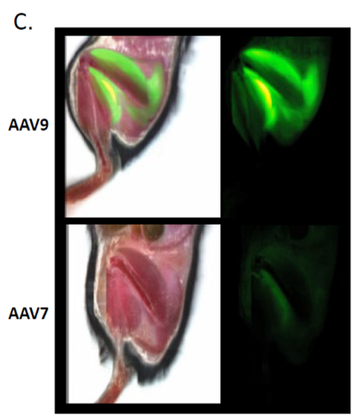
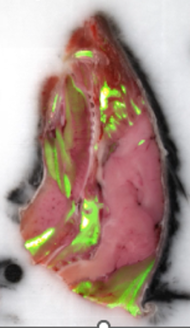
Example: Regional Transduction
AAV7-GFP + AAV9-tdTomato
Findings: CFT identified several tissues with AAV transduction and protein expression
Details:
Widespread transduction by both AAV7 (A) and AAV9 (B) in the muscle cells of the tongue, tissues in the sinus cavity, lacrimal and Harderian glands beneath the eye, and inside the marrow of the jaw and outside the roots of the incisors.
Yost et. al, ASGCT 2023