Video Gallery > Cryo-Fluorescence Tomography: Latest Insights in Preclinical Applications with Xerra
Cryo-Fluorescence Tomography: Latest Insights in Preclinical Applications with Xerra
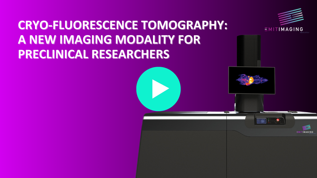
Cryo-Fluorescence Tomography (CFT) is a method to interrogate whole organs and animals for molecular probes, chiefly fluorescent markers, in 3D, at very high resolution and very high dynamic range. Xerra is a device from Emit Imaging which uses CFT to do tracking of gene expression, immunologics and early stage PK/PD and biodistribution studies.
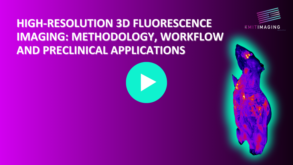
CFT is an ex vivo Molecular Tissue Imaging (MTI) technique where we generate 3D fluorescence and 3D anatomical images from individual 2D sections. CFT is a meso-scopic technique that can visualize cell clusters up to large organs within samples from isolated organs to whole intact animals and it is not exclusive of other techniques so fits into any research workflow. CFT is a powerful validation tool for invivo imaging, and can also facilitate more accurate histology and pathology studies like auto-rad, IHC, H&E etc. CFT uses LED laser excitation light with narrow band emission filters to produce a stack of 2D block face images to reconstruct into a 3D volume.