Introducing Emit Imaging
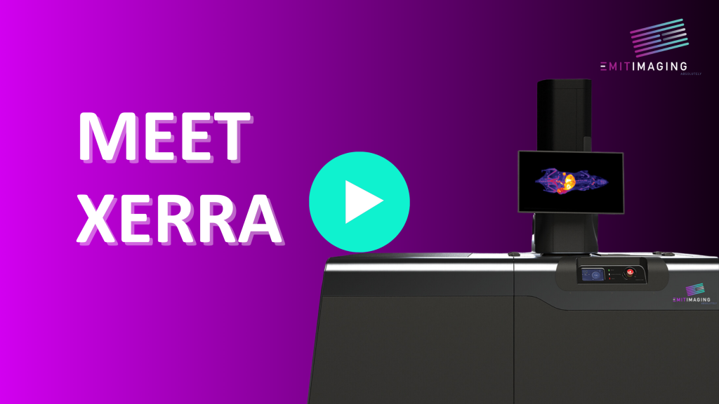
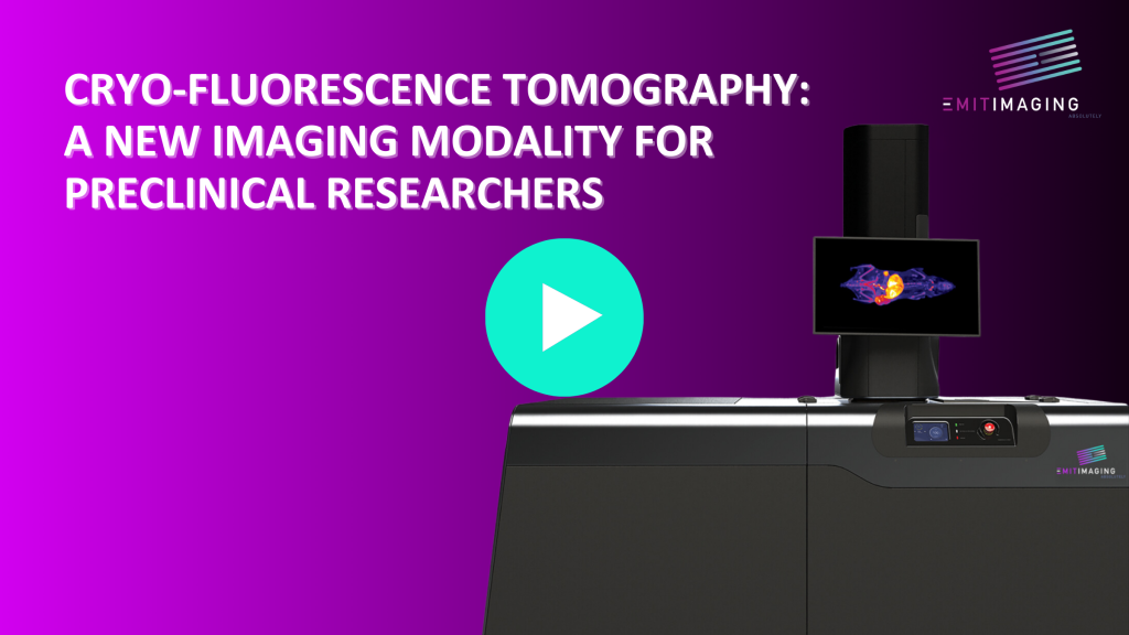
Meet Xerra, an important 3D tissue imaging technology developed by Emit Imaging. This preclinical imaging platform, is arguably the most disruptive imaging platform that has entered the market in the past 15 years. The last big players were bioluminescence, micro-PET, and high-frequency ultrasound, which have become indispensable ‘standards’ for preclinical research and drug development.
Preclinical researchers who depend on imaging have always struggled to get high resolution data especially from fluorescence imaging platforms. The Xerra resolves this historical struggle.
Xerra is the combination of a cryomacrotome and a multi-spectral optical imaging system enabling whole animal imaging of anatomy of any fluorophore at resolutions on the order of 20 microns.
The images are striking with potential to disrupt how we approach drug discovery and biodistribution studies. The pharmacokinetics/pharmacodynamics (ADME) category is critical and certainly financially attractive, but the Xerra offers unprecedented insights into numerous other applications including, but not limited to, neuroscience, cancer biology, immunology, cell, and nanoparticle tracking.


