
Antibody-drug conjugates
Gain insight into biodistribution, target engagement, and payload localization.
The imaging gap in ADC development
Antibody-drug conjugates are redefining targeted therapy by combining the specificity of antibodies with potent cytotoxic payloads. Despite their promise, many ADCs fail in translation due to incomplete understanding of how they distribute and interact in complex biological systems. Preclinical researchers are often faced with the following challenges when imaging their ADCs:
Given these challenges, researchers are left with gaps in spatial and molecular understanding that can lead to incorrect or incomplete conclusions about ADC targeting, safety, and efficacy.
EMIT Imaging's visualization tools make CFT data easier to explore and collaborate on, with a full range of options to fit different user types and use cases.
Contact us to identify the best visualization tool for your team and receive a demo →
Effective ADC design relies on clear visualization of both antibody targeting and payload delivery, enabling researchers to connect pharmacokinetics (PK), pharmacodynamics (PD), and therapeutic outcomes.
Image Gallery →Developing an ADC program?
Send us your study design and someone from our team will follow up.
Why is CFT essential for preclinical ADC research?
Conventional methods cannot provide a comprehensive, unbiased map of where an ADC travels and accumulates. Cryo-Fluorescence Tomography serves as a powerful technique by simultaneously providing broad spatial mapping along with high spatial resolution. CFT supports ADC researchers by:
By visualizing every tissue in 3D, researchers gain the anatomical and molecular context needed to thoroughly test ADC candidate design hypotheses and reduce preclinical uncertainty.
What is Cryo-Fluorescence Tomography?
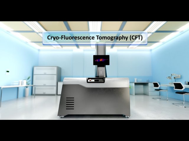
CFT is an ex vivo 3D imaging technique that captures both molecular fluorescence and anatomical data from entire animals or large tissue samples.
Cryo-embedding
Whole animals or tissues are frozen and embedded in a specialized media for imaging without chemical fixation.
Serial block-face imaging
The block face is repeatedly sectioned, with each newly exposed surface captured in fluorescence and white-light imaging.
3D reconstruction
The resulting images are aligned into a full volumetric dataset, combining molecular and anatomical detail.
How CFT integrates across the ADC development pipeline
CFT enables confident, data-driven decision-making throughout the preclinical workflow.
Partner with EMIT Imaging
Accelerate discovery and eliminate uncertainty with EMIT Imaging. Our Xerra™ instrumentation and expert CFT services deliver the same powerful combination of sensitivity and sub-organ-level resolution, giving you full-body visualization that traditional methods miss. Partnering with EMIT means faster insight, smarter study design, and more reliable data to drive your next breakthrough.
Want to see how your own data could look? Explore the full range of CFT visualization solutions.
Explore Visualization SolutionsExplore the data yourself, right in your browser
The EMIT Viewer is the web version of our visualization software. Open an example dataset to explore CFT for yourself, page through slices, toggle fluorescence channels, and rotate the reconstructed volume. Nothing to download and nothing to install.
Browse Example Datasets