Drug Discovery & Delivery
Monitor on- and off-target expression with resolution and sensitivity
Contact UsCFT for Drug Discovery and Delivery
Cryo-fluorescence tomography (CFT) is an ex vivo, volumetric tissue imaging technique used to monitor the pharmacokinetics and pharmacodynamics (PK/PD) of drugs and delivery vehicles with high-resolution and sensitivity. Images of the anatomy (white light) are acquired alongside fluorescence images that are displayed in a 3D visualization of the drug and/or delivery vehicle biodistribution.
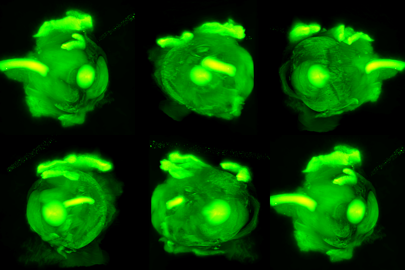
Example: AAV-Mediated Protein Expression
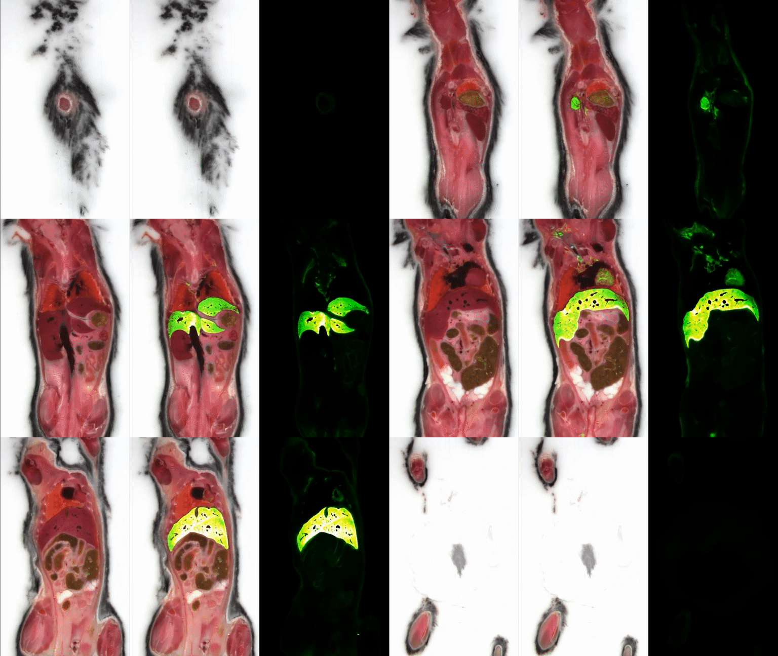
IV Administration
Findings: CFT uniquely achieves the required combination of resolution and sensitivity in a whole animal.
Details:
- GFP expression at day 15 following IV injection
- No expression in the control
- Liver is major organ where GFP expression occurs but protein expression is observed throughout the body
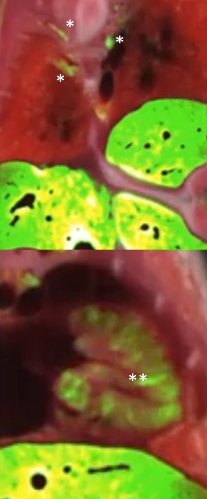
- Note signal in dorsal root ganglion (DRG) and heart, which is consistent with AAV9 tropism
Example: Visualization of Cell Intake
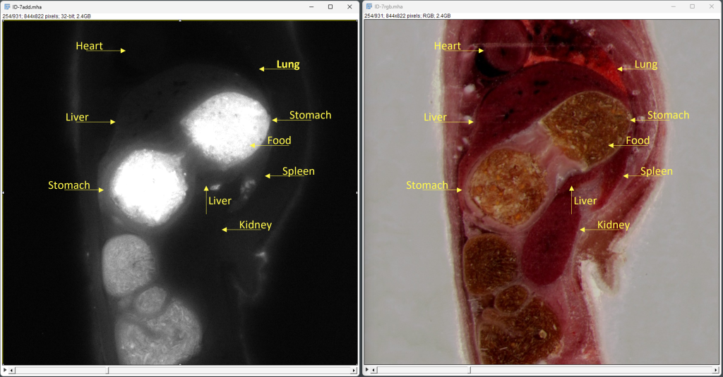
Intrasplenic Cell Injection
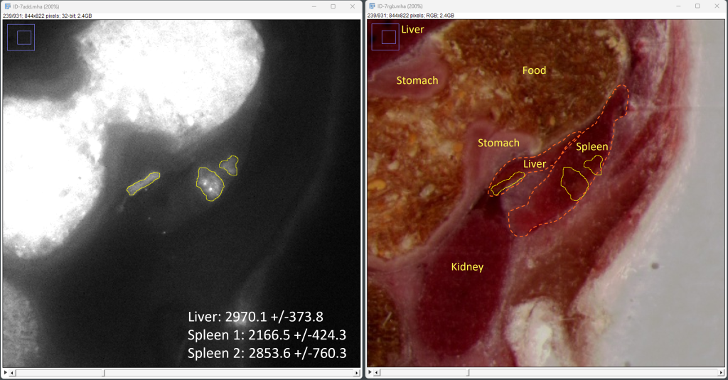
Findings: CFT was used to visualize labeled cell intake into tissue.
Details:
- Cell tracking by fluorescence labeling is a critical aspect of cell-based therapies, such as CAR-T and regenerative medicine.
- In preclinical research, cells are administered systemically or directly in target tissues, and localization is evaluated using in vivo or tissue imaging.
- In this example, proprietary cells were labeled with commercial QTracker 800 (ThermoFisher) and injected into mouse spleen.
- Primary pellet observed in the spleen and a secondary pellet in the liver
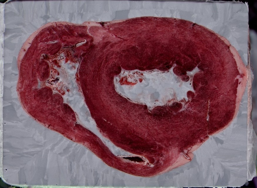
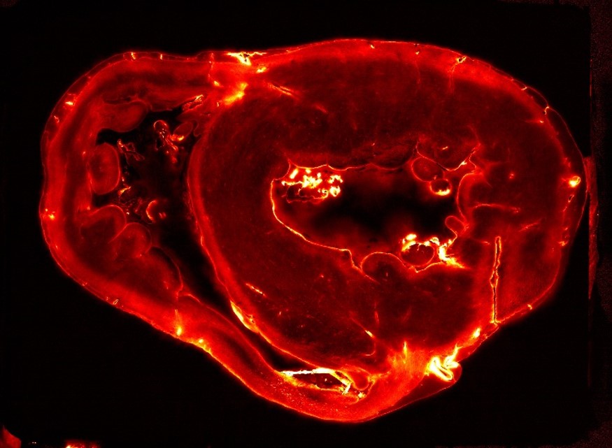
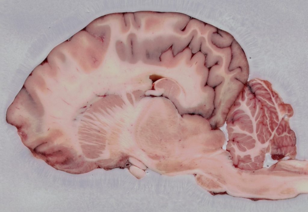
CFT Application in Large Animal Tissue
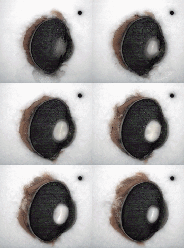
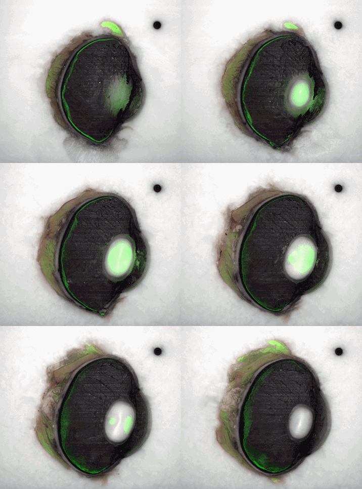
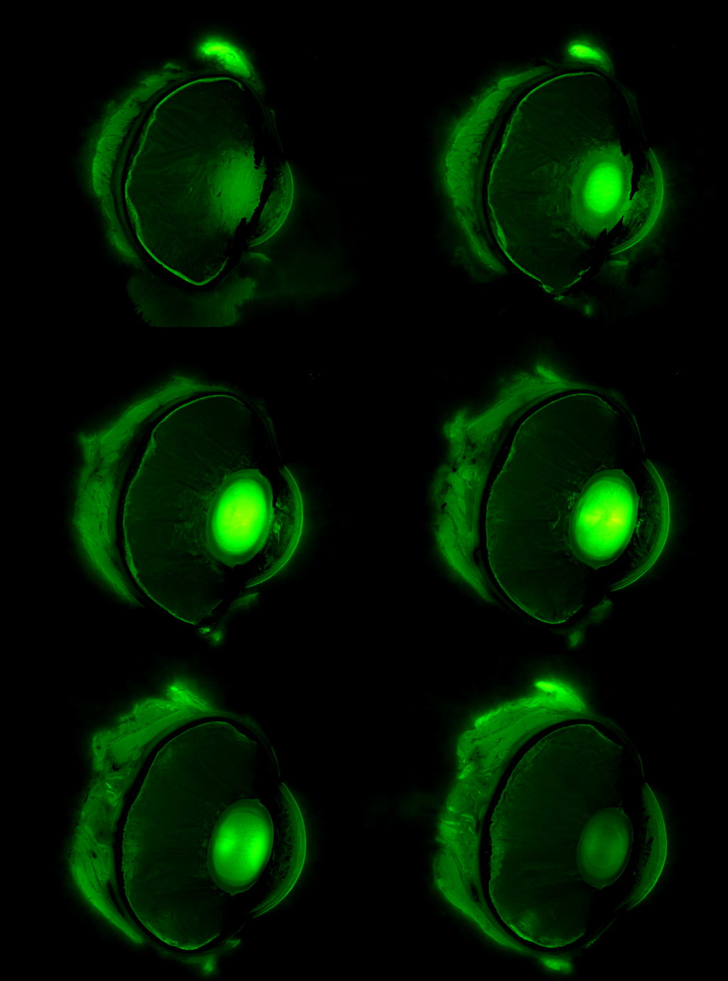
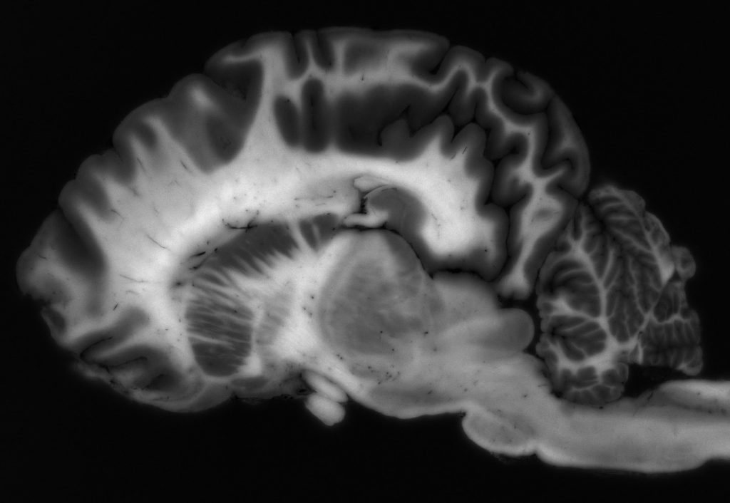
Because Xerra works in large formats, CFT has strong applications for anatomical RGB white-light and fluorescence imaging in large animal tissue, including NHP, Bovine, Porcine, and more.

