What is CFT?
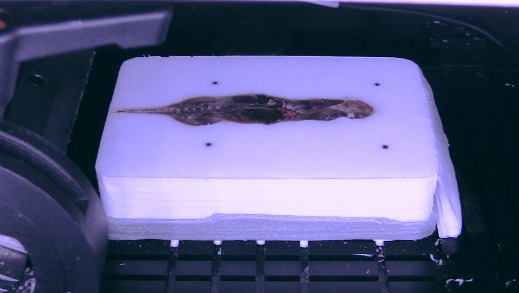
Cryo-Fluorescence Tomography (CFT) is an ex vivo, volumetric tissue imaging technique used to monitor the pharmacokinetics and pharmacodynamics (PK/PD) of drugs, delivery vehicles, and proteins with high resolution and sensitivity. White light, anatomical Images are acquired alongside fluorescence images that are displayed in a 3D visualization of the drug biodistribution and protein expression.
CFT is Complementary
Prior to CFT, an imaging gap existed between cellular microscopy and whole-body imaging modalities like MRI, PET, and CT. CFT is the bridge between cellular and whole-body imaging that could all be found on one platform.
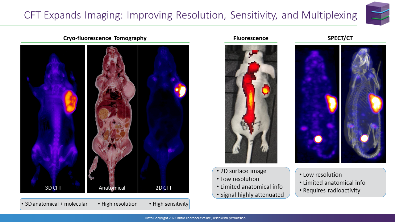
High-resolution and high-sensitivity CFT allows for detailed imaging of large tissue samples. In comparison, other imaging modalities can only focus on subcellular structures or low-resolution images of whole bodies or specific regions.
CFT Expands Imaging
Optical and Nuclear imaging methods are critical tools in research, however both have limitations. Standard, 2D optical imaging is easy to implement but is limited to low resolution, surface images with lacking anatomical information. Nuclear imaging techniques are 3D, but also provide low resolution with limited anatomical information, and require dedicated expertise and facilities that are impractical for some laboratories.
CFT increases the value of imaging studies by expanding the information provided. CFT can be used at key time points to describe PK/PD in more detail, providing both high-resolution anatomical and molecular information that improves the interpretation of standard optical and nuclear methods.
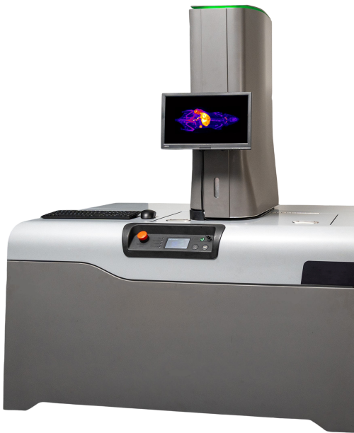
CFT Instrumentation & Services

Instrumentation

Services
EMIT Imaging offers CFT fee-for-service research performed in-house, at our state-of-the-art labs in Boston, MA and Baltimore, MD. Simply freeze samples, ship them directly to us, and let EMIT handle the rest.


