Preclinical Cryo-Fluorescence Tomography Imaging Services
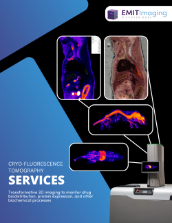
At EMIT Imaging, we offer advanced Cryo-Fluorescence Tomography (CFT) as a fee-for-service solution from our laboratory in Natick, Massachusetts. Our in-house experts guide you from study design through data analysis, delivering high-resolution, volumetric insights that support preclinical drug discovery and development.
CFT enables whole-animal and tissue imaging that connects molecular localization with anatomical context. By visualizing fluorescently labeled molecules in 3D, researchers can explore on- and off-target biodistribution, protein expression, and complex biochemical pathways with unprecedented clarity.
Whether you’re validating a gene therapy, characterizing a drug delivery vehicle, or tracking tumor progression, CFT delivers the section-by-section insights needed to make confident, data-driven decisions.

Get Started Today
Why Conduct a CFT Services Project?
Expertise From Study Design to Image Analysis
Visualize biodistribution, protein expression, and other processes in 3D with 20–55 µm resolution and single-digit nanomolar sensitivity.
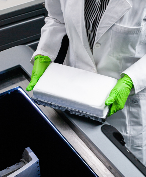
Work with our Project Managers and CFT experts from day one. We guide you through probe selection, food restrictions, and freezing protocols to ensure data quality from the start.
CFT can act as a screening tool ahead of downstream assays (IHC, LSM, biochemical analysis) or as the final step in a longitudinal study. We adapt to your study design.
Every CFT block is set up in a way that allows fluorescence units to be normalized utilizing the fluorescence units within blocks (standards) or across blocks (OCT). CFT users are able to analyze signal intensity across regions of interest.
After imaging, we identify and analyze key regions of interest (ROIs). Deliverables include raw and processed data, statistical summaries, and publication-ready visuals. Whether you need standard ROI analysis, more in-depth analysis, or something completely custom, EMIT Imaging’s team can accommodate. Each project generates 2D slice stacks, co-registered flythroughs, and 3D MIPs. While visually stunning, these images are designed to inform go/no-go decisions and strategic pivots. See more details on our typical Service Packages and deliverables in our Services Brochure here.
Each project is assigned a dedicated Project Manager to coordinate logistics and communication. Initial results are often delivered in as few as 14 business days, with final reports typically within 25 business days. For an example timeline and milestones associated with a service project, please click here to download our Services Brochure.
EMIT’s CFT services offer a cost-effective way to access the benefits of volumetric whole-body preclinical imaging without the upfront investment of purchasing a system. Ideal for low-volume projects or facilities with limited funding, it is an easily-accessible way to evaluate CFT before committing to in-house adoption. With EMIT’s fee-for-service CFT, you get the insights you need quickly, affordably, and backed by our expert support.


