Download The Brochure
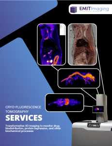
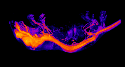
Cryo-Fluorescence Tomography is a volumetric tissue imaging technique used to visualize the 3D biodistribution of fluorescently-labeled molecules or the expression of fluorescent reporter proteins with high resolution and sensitivity. Anatomical and molecular images are co-registered, providing precise localization information of drugs, delivery vehicles, proteins, and other biochemical processes in whole animals and tissues.
Cryo-Fluorescence Tomography (CFT) has been widely adopted by researchers across industry and academia. The exceptional detail provided by CFT has proven critical in advancing research areas such as gene and cell therapy, drug discovery and delivery, oncology and immunotherapy, and neurology.
Cryo-Fluorescence Tomography fits seamlessly into existing workflows and, in many cases, can act as a replacement for in vivo optical imaging modalities. CFT provides higher resolution, higher sensitivity, and enhanced anatomical information compared to in vivo FLI, often correcting misinterpretations identified with other methods. Download the case study to learn more.
Speak with a member of our team by filling out the contact us form to schedule an introductory call