Adeno-Associated Virus Imaging
Cryo-Fluorescence Tomography (CFT) is a method to interrogate whole organs and animals for molecular probes, chiefly fluorescent markers, in 3D, at very high resolution and very high dynamic range. Xerra is a device from Emit Imaging which uses CFT to do tracking of gene expression, immunologics and early stage PK/PD and biodistribution studies. CFT is an ex vivo Molecular Tissue Imaging (MTI) technique where we generate 3D fluorescence and 3D anatomical images from individual 2D sections.
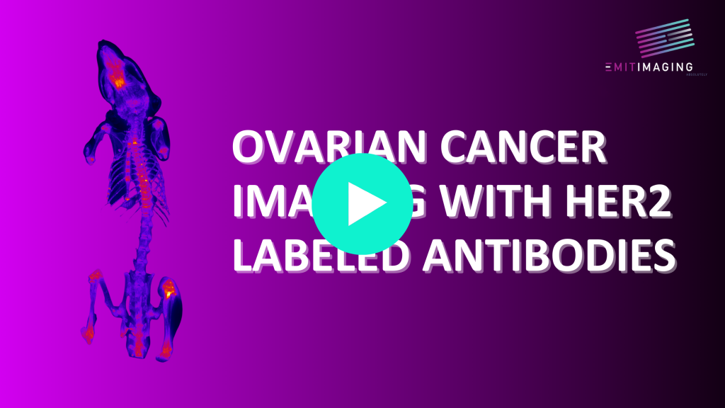
Adeno-associated virus vectors (AAV) are emerging tools in gene therapies, however, in their development, it can be very difficult to map the full extent of the gene delivery. CFT has been used to map Her2 expressing tumor cells which co-express Tdtomato, novel probes with a Her-2 protein targeting antibody labeled with Alexa Fluor IVIS data does not show non specific binding or renal clearance and is not good for longitudinal studies.