Macrophage Imaging
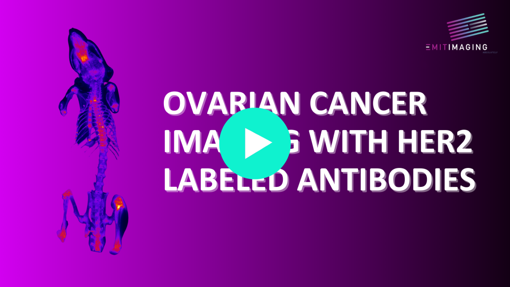
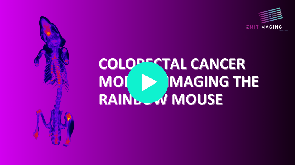
Cryo-Fluorescence Tomography (CFT) is a method to interrogate whole organs and animals for molecular probes, chiefly fluorescent markers, in 3D, at very high resolution and very high dynamic range. Xerra is a device from Emit Imaging which uses CFT to do tracking of gene expression, immunologics and early stage PK/PD and biodistribution studies. CFT is an ex vivo Molecular Tissue Imaging (MTI) technique where we generate 3D fluorescence and 3D anatomical images from individual 2D sections.
Xerra gives much better data on probes kinetics, targeting and non-specific binding. Researchers can label macrophages with a near infrared probe like Texas Red, Cy7 IRdye 800, IRdye 700, Cyanine based dyes, Rhodamine dyes, BODIPY dyes, Quantum Dots etc. to try to visualize inflammatory response. Macrophages and tumor associated macrophages in response to tumor growth are a great use of CFT. Xerra produces 16 bit images for large dynamic range and high resolution data. Macrophages can be seen in long bone and in the bone marrow along with circulation and heterogeneous uptake in the tumor and lymph nodes and liver and spleen.