Colorectal Cancer Models: Imaging the Rainbow Mouse
Cryo-Fluorescence Tomography (CFT) is a method to interrogate whole organs and animals for molecular probes, chiefly fluorescent markers, in 3D, at very high resolution and very high dynamic range. Xerra is a device from Emit Imaging which uses CFT to do tracking of gene expression, immunologics and early stage PK/PD and biodistribution studies. CFT is an ex vivo Molecular Tissue Imaging (MTI) technique where we generate 3D fluorescence and 3D anatomical images from individual 2D sections.
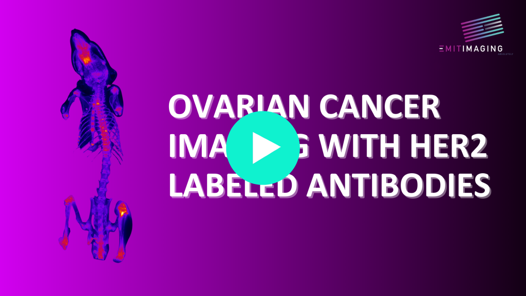
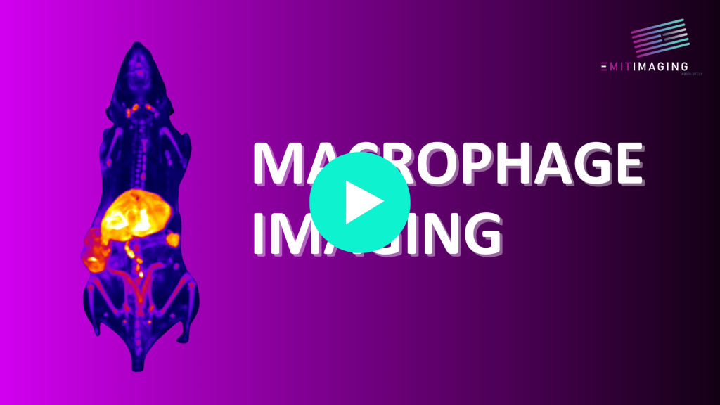
Xerra multi channel data gives us an excellent view of the tumor micro environment and can visualize Green Fluorescent Protein (GFP), Cerulean (CFP), Anthozoan fluorescent proteins, Yellow Fluorescent protein (YFP), Red fluorescent protein RFP, mCherry, TagRFP, etc. Fluorescent protein reporter genes in the Adeno associated virus vectors, AAVs, are a great application for CFT which can visualize AAV propagation through out the whole animal.
Since we are imaging the top surface we are not hindered by short wavelengths. 3D maximum intensity projections (MIPs) of Patient Derived Xenograft, PDx, tumors with a novel molecular probe injected IV prior to surgical resection can be imaged. Vascular images can be made with Indocyanine green (ICG), Evans Blue, Fluorescein and CFT can image Aminolevulinic Acid-Induced Protoporphyrin IX Fluorescence.