CFT Outperforms In Vivo FLI/BLI
Cryo-Fluorescence Tomography (CFT) is a transformative 3D approach to imaging drug distribution and protein expression (PK/PD) in whole animals. Compared to in vivo optical imaging techniques such as 2D fluorescence imaging (FLI) and bioluminescence imaging (BLI), CFT provides high resolution, high sensitivity anatomical and fluorescence images designed to help you discover more in biological and drug research. EMIT Imaging offers contract services and instrumentation (XerraTM), allowing for a seamless integration into your existing imaging workflows. See what you’re missing and utilize CFT to better visualize:
- Whole-body drug distribution and delivery
- Screening candidate drugs and delivery systems
- Whole-body therapeutic protein expression
- Multiplexed co-localization of drug with targets
- On and off target tissue identification

Download the full case study comparing the performance of Cryo-Fluorescence Tomography with 2D In Vivo Fluorescence
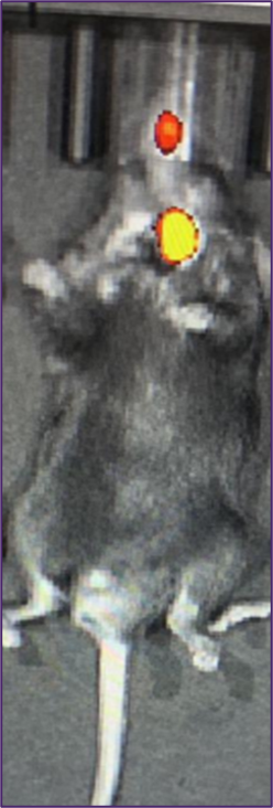
Cy-5.5 labeled Fibrin-Targeting Peptide (EP-2105)
Data provided by Junfeng Wang, Ph.D. | Harvard Medical School
Additional Resources


CFT vs. In Vivo FLI
In Vivo Imaging Provided Misleading Data in Fibrin-Targeting Drug Study
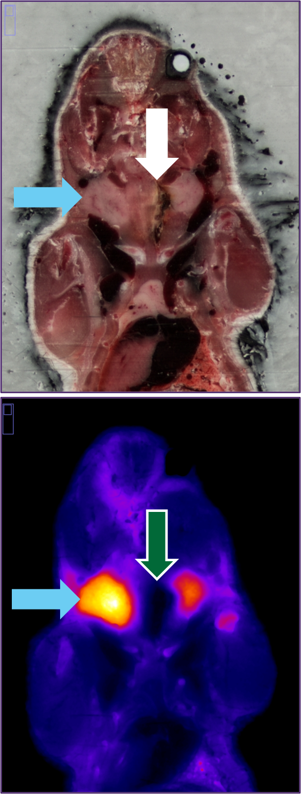
Findings: The lack of anatomical data and resolution from 2D in vivo imaging (IVIS) led to the incorrect interpretation that a fibrin-targeting peptide was localized to the thrombus in a FeCl3 thrombosis model. 3D CFT confirmed the presence of a thrombus in the anatomical image and showed that the fibrin-targeting peptide signal was in the submandibular gland, not the thrombus.
Details
- Ferric Chloride-induced murine thrombosis model (FeCl3)
- A Cy-5.5 labeled (BP-22460) fibrin- targeting peptide compound (EP-2105) was administered via tail vein injection
- Mice imaged 10 min post-injection
Data Provided By:
Junfeng Wang, PhD | Asst. Professor of Radiology
Gordon Center for Medical Imaging
Massachusetts General Hospital, Harvard Medical School
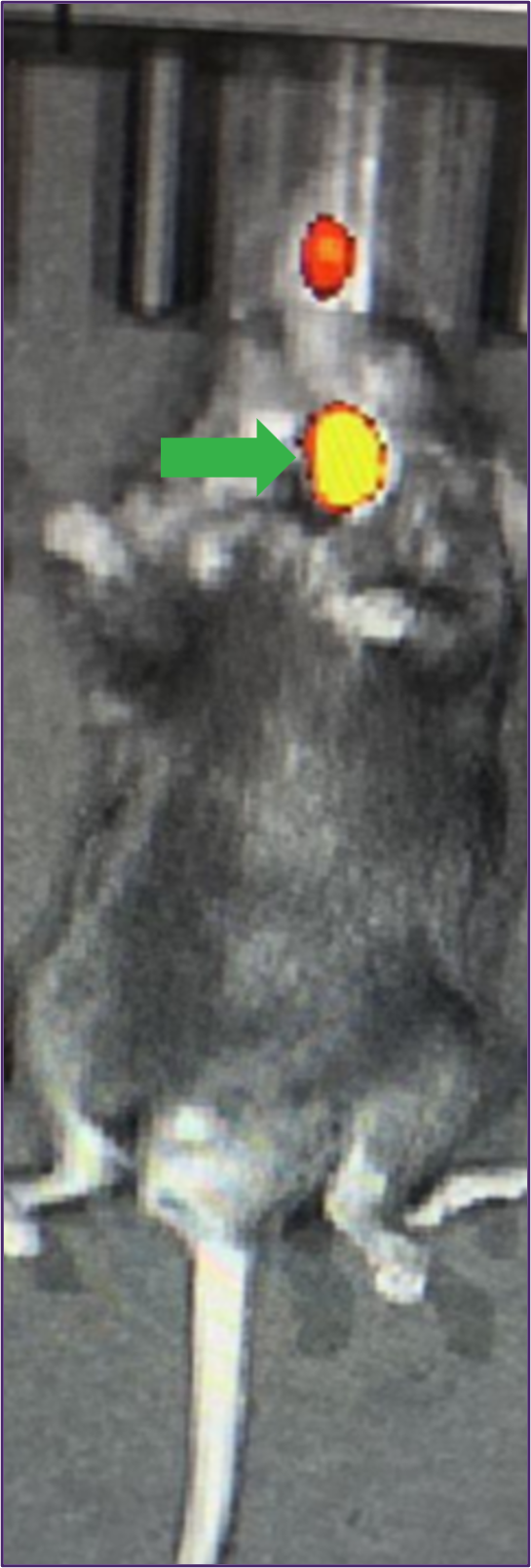
→ Signal interpreted as localization to thrombus from in vivo imaging
→ Signal shown as coming from the submandibular gland
→ Presence of thrombus was observed from high resolution anatomical CFT data
→ No accumulation of fluorescence-labeled peptide in the thrombus observed via CFT
High Sensitivity CFT Identified Signal In Thrombus where In Vivo Imaging Failed
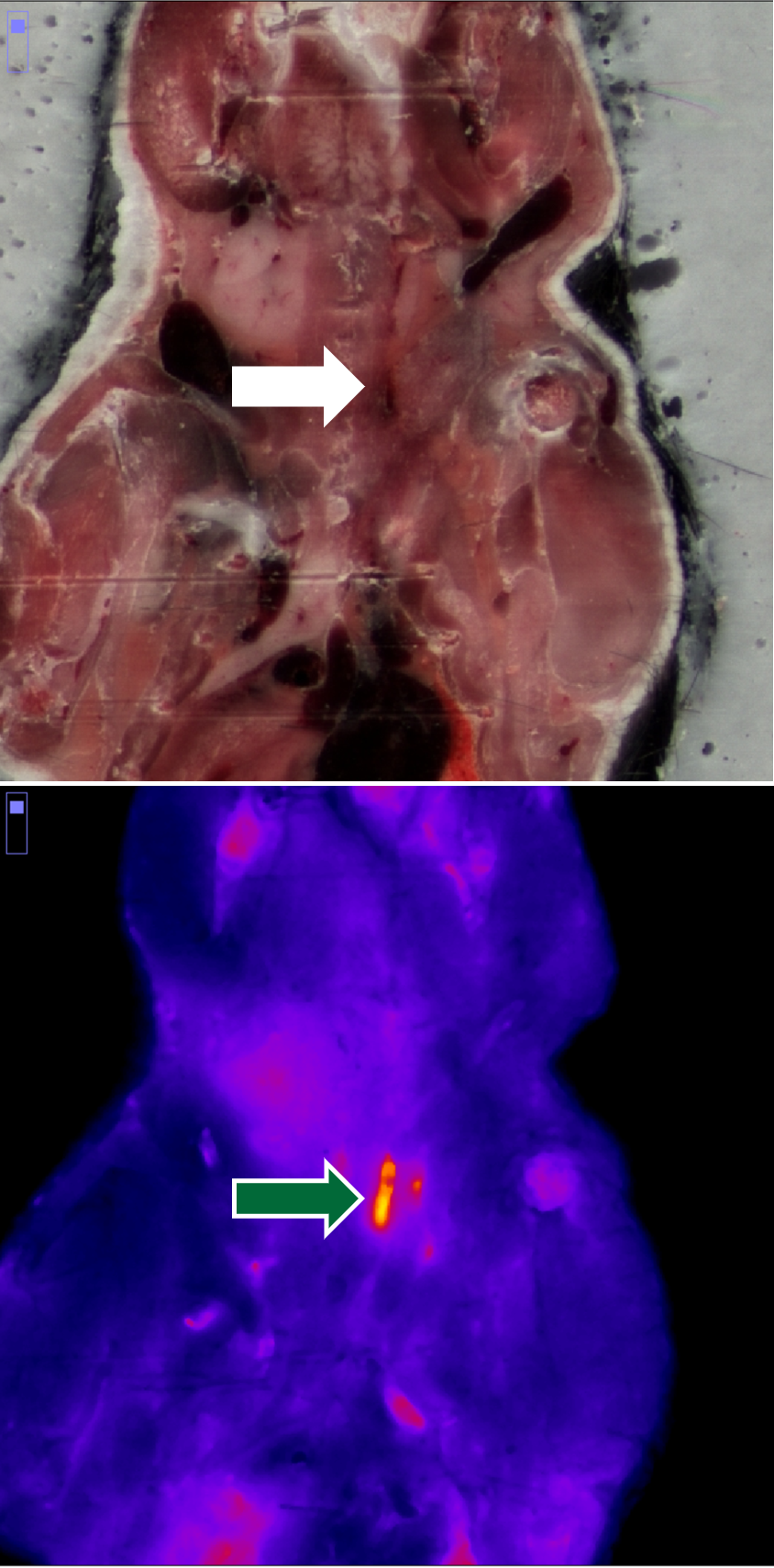
Findings: Due to the low sensitivity of in vivo imaging (IVIS), localization of a fibrin-targeting peptide to the thrombus was not observed in a vascular crush injury model. High resolution and sensitivity CFT identified the presence of the peptide in the thrombus as expected.
Details
- Vascular crush injury thrombosis model
- A Cy-5.5 labeled (BP-22460) fibrin- targeting peptide compound (EP-2105) was administered via tail vein injection
- Mice imaged 10 min post-injection
Data Provided By:
Junfeng Wang, PhD | Asst. Professor of Radiology
Gordon Center for Medical Imaging
Massachusetts General Hospital, Harvard Medical School
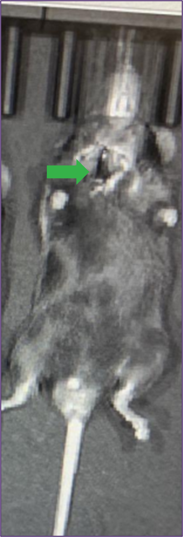
→ No signal/localization was observed in the thrombus with in vivo imaging
→ Presence of thrombus was observed from high resolution anatomical CFT data
→ Visualization of fibrin-targeting peptide to the thrombus observed via CFT
Multiplexing: Visualize 3D Co-localization of Drug and Tumor Cells
Findings: CFT shows high-resolution, 3D co-localization of tumor cells and drugs, complementing and expanding 2D in vivo imaging
Details
- Orthotopic ovarian cancer model with tdTomato-expressing cells
- Injected HER2-targeted antibody labeled with AF647
Ovarian Cancer cells (tdTomato)


Her2-Targeting Protein (AF647)
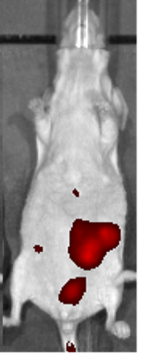
Intrathecal Injection: Whole Body Distribution of Labeled ASO
Findings: CFT is superior to surface fluorescence due to its depth limitations (signal loss) and poor resolution
Details:
- Cy5-labeled ASO at day 15 following IT injection
- Full visualization of spinal column into the cisterna magna
- CFT detects bioD and clearance: GI, lympathic, nasal turbinate, etc.
- CFT is 3D and high resolution: <50mm versus >1mm in optical and nuclear medicine imaging
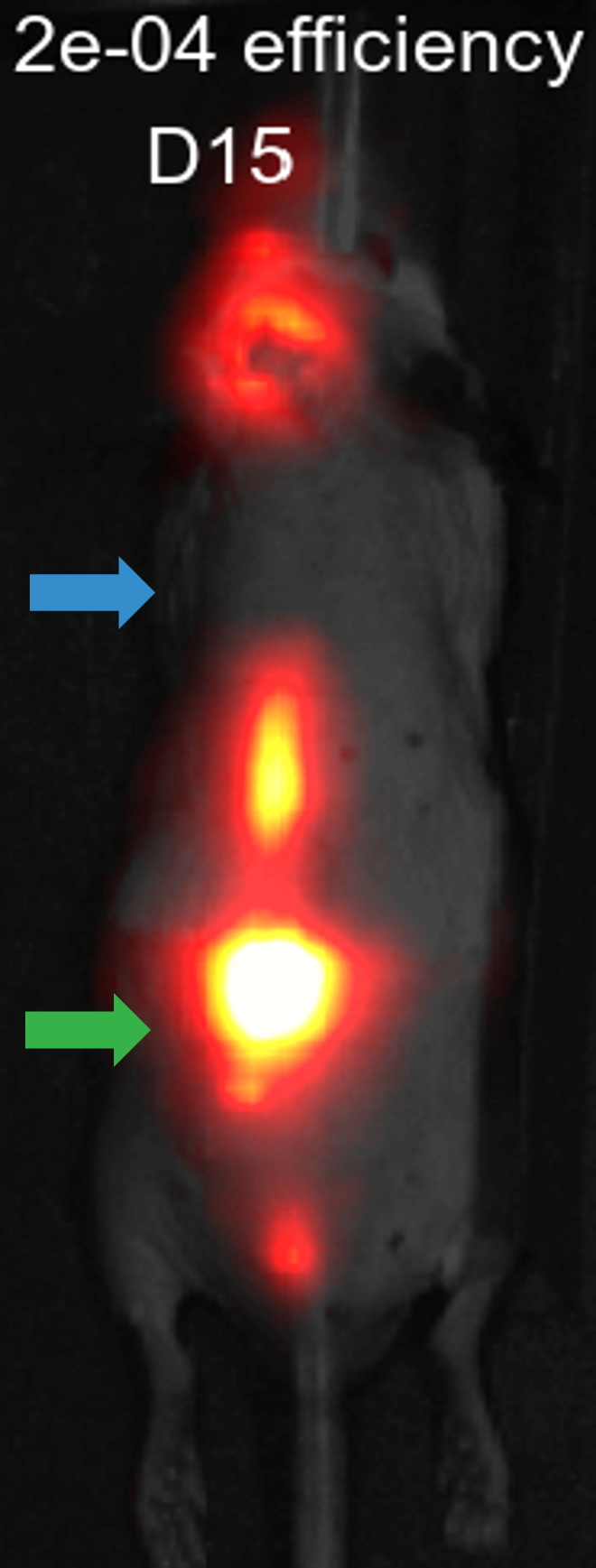
→ Signal loss due to depth, note the spine in CFT
→ Site of Injection
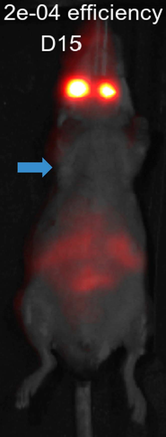
→Signal loss due to depth, note the spine in CFT
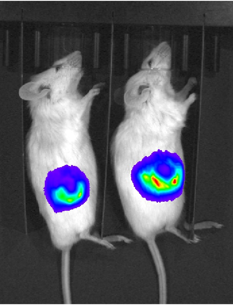
CFT vs. In Vivo BLI
Understanding of Disease Models
Findings: CFT image shows the primary tumor extent and heterogeneity as well as the metastatic lesions in the lungs
Details:
- MDA-MB-231 human breast adenocarcinoma cells were engineered to express both firefly luciferase and DsRed
- The in vivo bioluminescence image shows the flank tumor and can be used to track progression
DsRed mice provided by John Ronald, Robarts Research Institute
BLI (ffluc)
CFT (DsRed)